画像診断サービス
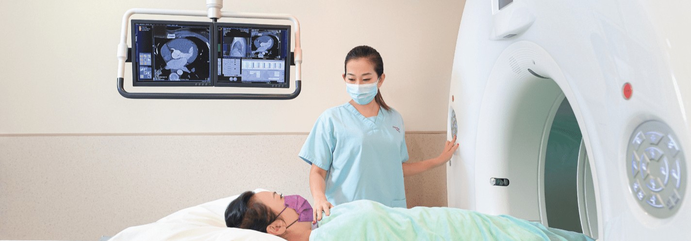
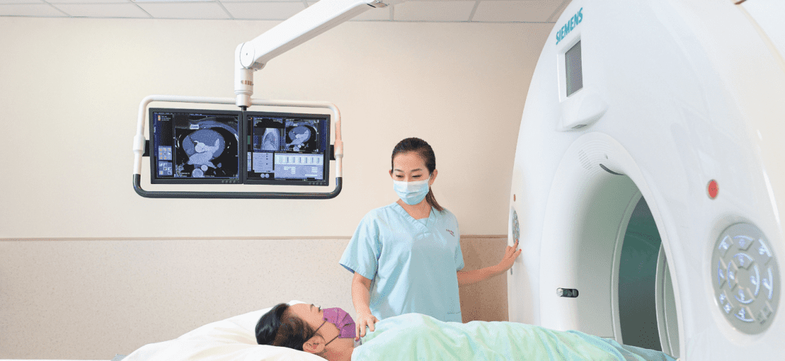
より正確な診断を下すためには、画像診断検査により体内の様々な臓器の形態や機能を調べることが大変有効です。
香港アドベンティスト病院の画像診断部には、精密な画像診断検査を迅速に行うための最先端医療設備が整っており、この分野の専門をする放射線科医/検査技師によって検査が行われています。
当院では、患者第一の理念のもと、外来/入院患者のために様々な画像診断検査を提供しています。
Magnetic Resonance Imaging (MRI)
MRI stands for Magnetic Resonance Imaging. Unlike X-rays and CT Scans, MRI is a completely non-invasive imaging technique and does not contain radiation. The magnetic field produced by an MRI forces certain atoms in your body to line up in a particular way. Radio waves are sent towards these atoms and bounce back, and a computer records the signal produced. Since different types of tissues send back different signals, the computer can analyze the signals and use them to render a detailed image of the internal organs.
MAGNETOM Skyrafit, features Siemens’ new generation MRI technology. The 70-cm open bore design carries a load of up to 250 kg and works well for claustrophobic patients and those with special needs. At 173 cm long, the system also allows for the head of the patient to remain outside the bore during scanning,
Our state-of-the-art MRI equipment can provide the following services:
- Brain MRI Scan
- Spine MRI Scan
- Cervical, Thoracic and Lumbar Spine Scans
- Cardiac MRI (Heart) Scans
- MRA (Magnetic Resonance Angiography)
- Other High Resolution Regional MRI Scans
- Musculoskeletal and Joint imaging
- MR Cholangiogram
- Abdominal and Chest
Computed Tomography (CT Scan)
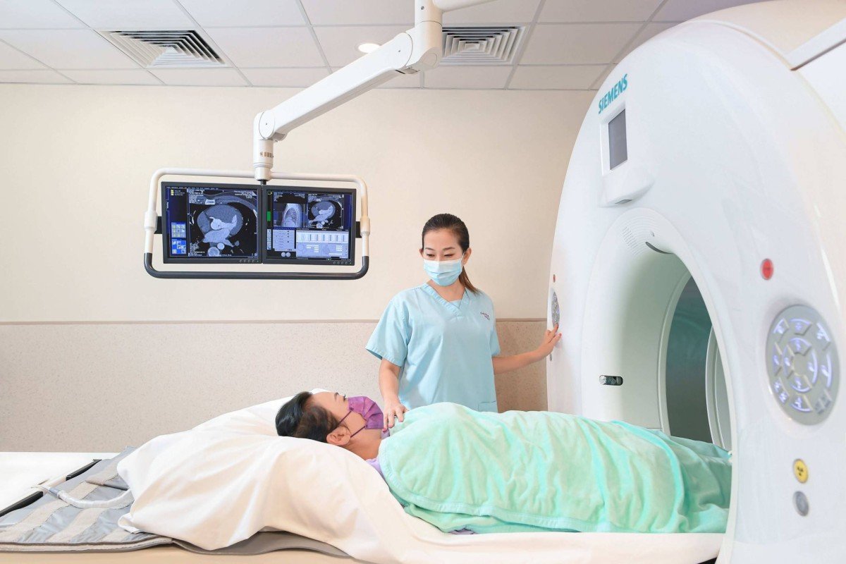
CT (Computerized Tomography) Scan, is the use of computer technology to translate our “flat” X-ray image into slices of images. Through study of hundreds of images and the application of 3D and 4D-display functions, 3D volumetric information of the body can be obtained in CT Scan. The CT computer also allows us to enhance the contrast of the displayed tissues, therefore we can pick up subtle change in soft tissue to identify pathology in the earliest possible stage. The X-ray characteristics and its high scanning speed make CT Scan ideal for abdominal, thorax, bony, and traumatic imaging.
Our high-speed Spiral CT Scan also allows us to perform high resolution CT Angiogram. CT Angiogram is commonly used as an initial investigation of suspected Arterial Vessel disease and it significantly reduces the number of Conventional Angiogram. At the same time, it also decreases the cost and the risk of Angiogram.
Siemens SOMATOM Definition Flash 256-slices CT Scanner has a dual-source and dual-energy design featuring two X-ray tubes that simultaneously revolve around the patient’s body. The design enables better tissue differentiation and a reduction of the number of scans.
Mammogram
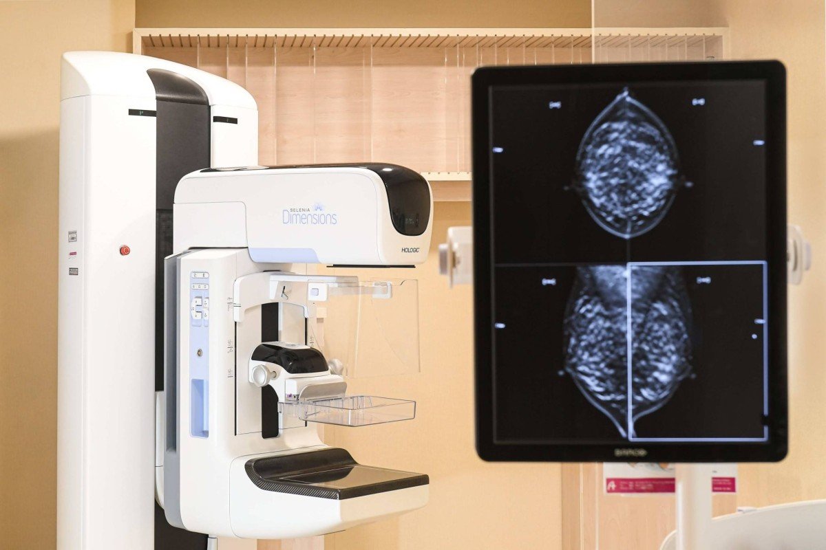
A mammogram will allow doctors to detect any abnormal breast tissue, or untouched/unformed tumor locations (calcification points), which may be a sign of early stage breast cancer.
Our Hospital offers:
- 2D Mammogram
- 3D Mammogram
一般Ⅹ線撮影およびX線透視検査
Radiology, otherwise known as X-ray, is the use of high penetration radiation to produce 2D picture of our 3D body tissue on film for diagnostic purpose. Radiology is the most common and relatively inexpensive imaging modality used in Diagnostic Imaging. Radiology is good for tissue of high inherent contrast such as bone and lung.
Our X-Ray department provides the following services for both in and out patients:
- General X-Ray, such as chest and bone X-ray.
- Barium Meal - Examine the upper alimentary system and stomach.
- Barium Enema - Examination of colon.
- Intravenous Pyelogram or IVP - Examination of the urinary system for possible obstruction, infection or mass within the system.
- Hysterosalpingography or HSG - Examination of the female reproduction system and is commonly used for investigation of infertility.
- Venography - For the examination of the venous system in the body for deep vein thrombosis.
- Biopsy for pathology
骨密度検査(DEXA)
Our Hospital is equipped with the GE Lunar Prodigy ADVANCE dual-energy bone densitometer, which has a wider scanning range and can scan the whole body for both bone density and body composition at once.
With the use of 2 different penetration power of X-ray, DEXA is used to measure the density of our bones (calcium and other minerals). With the X-ray dose of lesser than a chest X-ray and relatively safe, DEXA is commonly used to measure bone density of Spine, Hip, Wrist, and the most frequently fracture locations.
Body composition refers to the content and composition ration of fat, lean tissue, and bone minerals in the total mass of the human body. Compared to other examinations, DEXA has the advantages of shorter scanning time, higher precision, and higher accuracy.
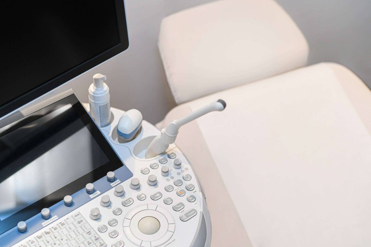
超音波検査
体に超音波プローブを当て、体内の臓器から跳ね返ってきた超音波を画像として映し出すことで、臓器の構造や形態に異常がないかを調べます。被爆の心配がないため、妊娠中の女性にも幅広く行われています。
Positron Emission Tomography-Computed Tomography (PET-CT)
Positron Emission Tomography-Computed Tomography (PET-CT) is a combined examination of both cellular structures and functions of the body. Apart from detecting pathological changes of tumour cells in the early stages and assessing the extent of metastasis, PET-CT scan can also be used to diagnose cardiovascular and neurological diseases, such as primary brain tumour, Alzheimer’s diseases and seizure.
Clinical applications of PET-CT includes:
Clinical applications of PET-CT includes:
- Diagnosing cancer at an early stage
- Identifying the site of primary cancer
- Localizing the site for biopsy
- Facilitating tumour grading and staging
- Identifying metastasis if any
- Formulating a treatment plan such as radiation therapy, chemotherapy
- Evaluating patient’s prognosis
- Assessing the effectiveness of treatment
- Monitoring cancer recurrence if any
Radionuclide & Molecular Imaging (RNMI)
Radionuclide & Molecular Imaging diagnoses and cures diseases using radiopharmaceuticals. The scanning tests use a special camera to take pictures of certain tissues in the body for clinical diagnosis after a radioactive tracer (radiopharmaceutical) is given to the patient. The tracer accumulates in the tissues, making them visible on the camera. Its extensive application ranges from tumors, brain, heart, lungs, the alimentary canal, bones, endocrine to urinary system and so on. It can effectively detect early expansion of tumors and physiological disorder of organs.
Environment
I hope this message finds you well. My name is Samantha, and I would like to take a moment to express my heartfelt gratitude for the exceptional care I received at the Radiation Therapy Department of Hong Kong Adventist Hospital from December 2 to December 20, 2024.
During my 15 sessions of treatment, I was fortunate to be under the care of three outstanding staff members: Adrean, Fai, and Jancy. Their professionalism, compassion, and dedication made a significant difference in my experience. They not only provided excellent medical care but also offered emotional support that helped me through a challenging time.
On my last day of treatment, which coincided with my birthday, Adrean, Fai, and Jancy surprised me by playing a birthday song. This thoughtful gesture truly touched my heart and made my day extra special. It is this kind of personalized care that sets your hospital apart.
As the holiday season approaches, I wish everyone at the hospital a joyful and peaceful time. Thank you once again for your outstanding service and for having such wonderful team members.
お問い合わせ: 28350515
DIAGNOSTIC IMAGING DEPARTMENT
|
Items |
項目 |
VIP/Private |
Semi-Private |
Standard |
Out-Patient |
|
HK$ |
HK$ |
HK$ |
HK$ |
||
|
Computed Tomography (CT Scan) |
電腦掃描 |
|
|
|
|
|
CT brain (without contrast) |
腦部(平片) |
$5,080 |
$4,400 |
$3,350 |
$2,750 |
|
CT brain (W/WO Contrast) |
腦部(單用顯影劑) |
$9,150 |
$7,920 |
$6,030 |
$4,950 |
|
CT Thorax (W/WO Contrast) |
胸部(平片及顯影劑) |
$13,020 |
$11,260 |
$8,580 |
$7,040 |
|
CT Upper Abdomen (W/WO Contrast) |
上腹(平片及顯影劑) |
$12,610 |
$10,910 |
$8,320 |
$6,820 |
|
CT Lumbar Spine |
腰椎 |
$8,750 |
$7,560 |
$5,770 |
$4,730 |
|
CT Upper Extremity: Each Area No Contrast |
上肢: 一個部位(平片) |
$7,520 |
$6,510 |
$4,960 |
$4,070 |
|
CT Lower Extremity: Each Area No Contrast |
下肢: 一個部位(平片) |
$7,520 |
$6,510 |
$4,960 |
$4,070 |
|
Magnetic Resonance Imaging (MRI) |
磁力共振 |
||||
|
MR Brain (Without Contrast) |
腦掃描(平片) |
$13,460 |
$11,640 |
$8,880 |
$7,280 |
|
MR Brain (W/WO Contrast) |
腦掃描(平片及顯影劑) |
$19,890 |
$17,200 |
$13,110 |
$10,752 |
|
MR Liver (With Contrast) |
肝臟掃描(平片及顯影劑) |
$22,370 |
$19,350 |
$14,750 |
$12,096 |
|
MR Pelvis (With Contrast) |
盆骨腔器官掃描(平片及顯影劑) |
$22,370 |
$19,350 |
$14,750 |
$12,096 |
|
MR Lumbar Spine (Without Contrast) |
腰椎(平片) |
$12,780 |
$11,050 |
$8,430 |
$6,912 |
|
MR Extremity (1 Region) (No Contrast) |
上或下支掃描(平片) |
$13,260 |
$11,460 |
$8,740 |
$7,168 |
|
Radionuclide & Molecular Imaging |
放射同位素診斷 |
||||
|
NM Bone Scan |
骨骼掃描 |
$16,200 |
$14,010 |
$11,210 |
$8,760 |
|
NM Myocardial Perfusion Scan (Rx Stress) |
心肌灌注雙核素掃描- 藥物輔助 |
$26,820 |
$23,200 |
$18,560 |
$14,500 |
|
X-Ray |
X光 |
|
|
|
|
|
XR Skull - Single Lateral view |
頭顱 -側位 |
$690 |
$600 |
$520 |
$378 |
|
XR Chest - Single view (Routine) |
胸部 (1像) |
$710 |
$620 |
$540 |
$389 |
|
XR Mammogram |
乳房造影 (雙側) |
$2,890 |
$2,500 |
$2,190 |
$1,565 |
|
XR KUB (Routine) - Single view |
泌尿系統平片 |
$750 |
$650 |
$570 |
$410 |
|
XR Cervical Spine - AP & Lat |
頸椎 (正位及側位) -2像 |
$1,340 |
$1,160 |
$1,010 |
$725 |
|
XR LS Spine - AP & Lat (routine) |
腰椎 (2像) |
$1,340 |
$1,160 |
$1,010 |
$725 |
|
XR Extremities - Two views |
上肢或下肢 -2像 |
$1,100 |
$950 |
$830 |
$599 |
|
XR Barium Meal |
胃及十二指腸鋇餐造影 |
$3,490 |
$3,020 |
$2,410 |
$1,890 |
|
XR Barium Enema |
大腸鋇劑灌腸造影 |
$6,210 |
$5,370 |
$4,300 |
$3,360 |
|
Ultrasound (US) |
超聲波 |
|
|
|
|
|
US Thyroid |
甲狀腺 |
$3,010 |
$2,600 |
$1,980 |
$1,628 |
|
US Upp Abd: GB, Liver ,Kid ,Pancreas ,Spleen |
上腹部(肝、膽、胰、脾) |
$5,050 |
$4,360 |
$3,330 |
$2,730 |
|
US Pelvis |
婦科 |
$3,010 |
$2,600 |
$1,980 |
$1,628 |
|
PET-CT |
正電子及電腦雙融掃描 |
|
|
|
|
|
PET / CT Whole Body Trunk Survey |
全身正電子及電腦掃描雙融掃描 |
$31,430 |
$20,280 |
$15,600 |
$15,600 |
|
PET / CT Whole Body Trunk Survey (+C) |
全身正電子及電腦掃描雙融掃描 (CK) |
$35,460 |
$22,880 |
$17,600 |
$17,600 |
|
Room Type |
||
|
VIP or Private: Single |
Semi-Private: Single or 2 Beds |
Standard Ward: 3 or 5 Beds |
Remarks:
- The above prices are for your reference. There may be further details that are not included here. Please re-confirm the prices with our staff prior to receiving treatments or examinations.
- Charges will be adjusted for urgent care service and services provided during non-office and non-clinic hours.
- Effective Date:2025/1/1(Subject to the latest version)
WeChat account name: hkahonc
By appointment
24時間救急医療サービス



.jpg)


